5.4 Errors in Meiosis
By the end of this section, you will be able to:
- Explain how nondisjunction leads to disorders in chromosome number
- Describe how errors in chromosome structure occur through inversions and translocations
Inherited disorders can arise when chromosomes behave abnormally during meiosis. Chromosome disorders can be divided into two categories: abnormalities in chromosome number and chromosome structural rearrangements. Because even small segments of chromosomes can span many genes, chromosomal disorders are characteristically dramatic and often fatal.
Disorders in Chromosome Number
The isolation and microscopic observation of chromosomes form the basis of cytogenetics and is the primary method by which clinicians detect chromosomal abnormalities in humans. A karyotype is the number and appearance of chromosomes, including their length, banding pattern, and centromere position. To obtain a view of an individual’s karyotype, cytologists photograph the chromosomes and then cut and paste each chromosome into a chart or karyogram (Figure 5.4.1).

Geneticists Use Karyograms to Identify Chromosomal Aberrations
The karyotype is a method by which traits characterized by chromosomal abnormalities can be identified from a single cell. To observe an individual’s karyotype, a person’s cells (like white blood cells) are first collected from a blood sample or other tissue. In the laboratory, the isolated cells are stimulated to begin actively dividing. A chemical is then applied to the cells to arrest mitosis during metaphase. The cells are then fixed to a slide.
The geneticist then stains chromosomes with one of several dyes to better visualise the distinct and reproducible banding patterns of each chromosome pair. Following staining, chromosomes are viewed using bright-field microscopy. An experienced cytogeneticist can identify each band. In addition to the banding patterns, chromosomes are further identified on the basis of size and centromere location. To obtain the classic depiction of the karyotype in which homologous pairs of chromosomes are aligned in numerical order from longest to shortest, the geneticist obtains a digital image, identifies each chromosome, and manually arranges the chromosomes into this pattern.
At its most basic, the karyogram may reveal genetic abnormalities in which an individual has too many or too few chromosomes per cell. Examples of this are Down syndrome, which is identified by a third copy of chromosome 21, and Turner syndrome, which is characterized by the presence of only one X chromosome in women instead of two. Geneticists can also identify large deletions or insertions of DNA. For instance, Jacobsen syndrome, which involves distinctive facial features as well as heart and bleeding defects, is identified by a deletion on chromosome 11. Finally, the karyotype can pinpoint translocations, which occur when a segment of genetic material breaks from one chromosome and reattaches to another chromosome or to a different part of the same chromosome. Translocations are implicated in certain cancers, including chronic myelogenous leukemia.
By observing a karyogram, geneticists can actually visualise the chromosomal composition of an individual to confirm or predict genetic abnormalities in offspring even before birth.
Nondisjunctions, Duplications and Deletions
Of all the chromosomal disorders, abnormalities in chromosome number are the most easily identifiable from a karyogram. Disorders of chromosome number include the duplication or loss of entire chromosomes, as well as changes in the number of complete sets of chromosomes. They are caused by nondisjunction, which occurs when pairs of homologous chromosomes or sister chromatids fail to separate during meiosis. The risk of nondisjunction increases with the age of the parents.
Nondisjunction can occur during either meiosis I or II, with different results (Figure 5.4.2). If homologous chromosomes fail to separate during meiosis I, the result is two gametes that lack that chromosome and two gametes with two copies of the chromosome. If sister chromatids fail to separate during meiosis II, the result is one gamete that lacks that chromosome, two normal gametes with one copy of the chromosome, and one gamete with two copies of the chromosome.
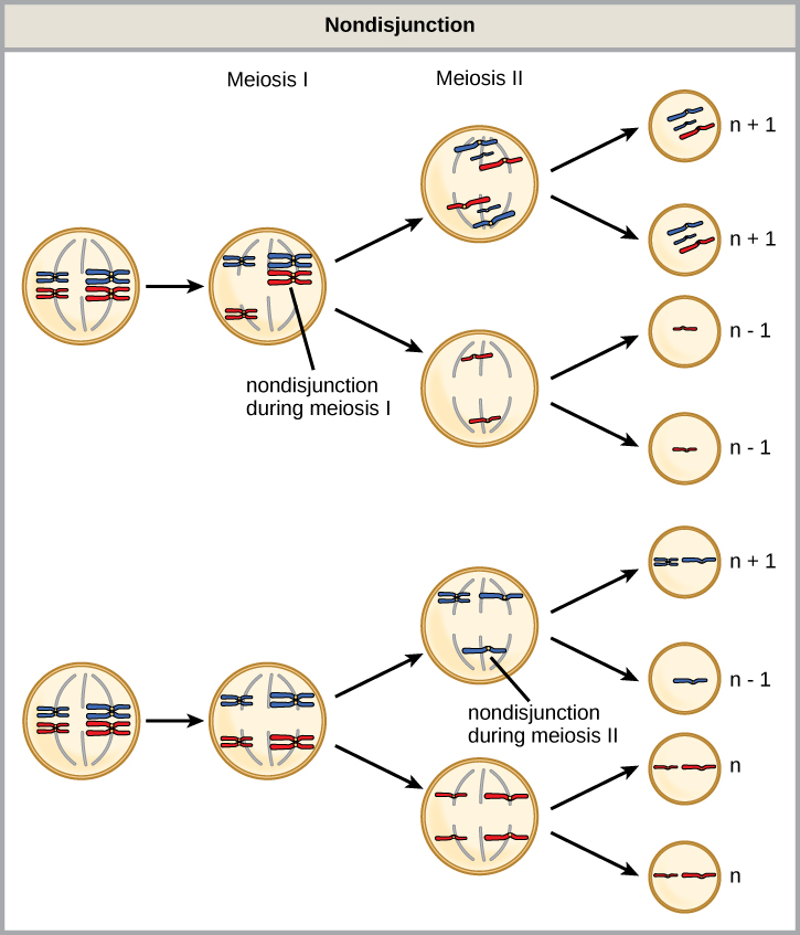
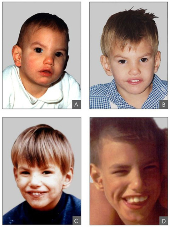
An individual with the appropriate number of chromosomes for their species is called euploid; in humans, euploidy corresponds to 22 pairs of autosomes and one pair of sex chromosomes. An individual with an error in chromosome number is described as aneuploid, a term that includes monosomy (loss of one chromosome) or trisomy (gain of an extraneous chromosome). Monosomic human zygotes missing any one copy of an autosome invariably fail to develop to birth because they have only one copy of essential genes. Most autosomal trisomies also fail to develop to birth; however, duplications of some of the smaller chromosomes (13, 15, 18, 21, or 22) can result in offspring that survive for several weeks to many years. Trisomic individuals suffer from a different type of genetic imbalance: an excess in gene dose. Cell functions are calibrated to the amount of gene product produced by two copies (doses) of each gene; adding a third copy (dose) disrupts this balance. The most common trisomy is that of chromosome 21, which leads to Down syndrome. Individuals with this inherited disorder have characteristic physical features and developmental delays in growth and cognition. The incidence of Down syndrome is correlated with maternal age, such that older women are more likely to give birth to children with Down syndrome (Figure 5.4.3).
Humans display dramatic deleterious effects with autosomal trisomies and monosomies. Therefore, it may seem counterintuitive that human females and males can function normally despite carrying different numbers of the X chromosome. In part, this occurs because of a process called X inactivation. Early in development, when female mammalian embryos consist of just a few thousand cells, one X chromosome in each cell inactivates by condensing into a structure called a Barr body. The genes on the inactive X chromosome are not expressed. The particular X chromosome (maternally or paternally derived) that is inactivated in each cell is random, but once the inactivation occurs, all cells descended from that cell will have the same inactive X chromosome. By this process, females compensate for their double genetic dose of the X chromosome.
Several errors in sex chromosome numbers have been characterized. Individuals with three X chromosomes, called triplo-X, appear female but express developmental delays and reduced fertility. The XXY chromosome complement, corresponding to one type of Klinefelter syndrome, corresponds to male individuals with small testes, enlarged breasts, and reduced body hair. The extra X chromosome undergoes inactivation to compensate for the excess genetic dosage. Turner syndrome, characterized as an X0 chromosome complement (i.e., only a single sex chromosome), corresponds to a female individual with short stature, webbed skin in the neck region, hearing and cardiac impairments, and sterility.
Chromosome Structural Rearrangements
Cytologists have characterized numerous structural rearrangements in chromosomes, including partial duplications, deletions, inversions, and translocations. Duplications and deletions often produce offspring that survive but exhibit physical and mental abnormalities. Cri-du-chat (from the French for “cry of the cat”) is a syndrome associated with nervous system abnormalities and identifiable physical features that result from a deletion of most of the small arm of chromosome 5 (Figure 5.4.3). Infants with this genotype emit a characteristic high-pitched cry upon which the disorder’s name is based.
Chromosome inversions and translocations can be identified by observing cells during meiosis because homologous chromosomes with a rearrangement in one of the pairs must contort to maintain appropriate gene alignment and pair effectively during prophase I.
A chromosome inversion is the detachment, 180° rotation, and reinsertion of part of a chromosome. Unless they disrupt a gene sequence, inversions only change the orientation of genes and are likely to have milder effects than aneuploid errors.
Section Summary
- The number, size, shape, and banding pattern of chromosomes make them easily identifiable in a karyogram and allow for the assessment of many chromosomal abnormalities.
- Disorders in chromosome number, or aneuploidies, are typically lethal to the embryo, although a few trisomic genotypes are viable.
- Chromosome structures may also be rearranged, for example, by inversion or translocation. Both of these aberrations can result in negative effects on development or death. Because they force chromosomes to assume contorted pairings during meiosis I, inversions and translocations are often associated with reduced fertility because of the likelihood of nondisjunction.
the number and appearance of an individuals chromosomes, including the size, banding patterns, and centromere position
the photographic image of a karyotype
the process by which one segment of a chromosome dissociates and reattaches to a different, nonhomologous chromosome
the failure of synapsed homologs to completely separate and migrate to separate poles during the first cell division of meiosis
an individual with the appropriate number of chromosomes for their species
an individual with an error in chromosome number; includes deletions and duplications of chromosome segments
an otherwise diploid genotype in which one chromosome is missing
an otherwise diploid genotype in which one entire chromosome is duplicated
any of the non-sex chromosomes
the condensation of X chromosomes into Barr bodies during embryonic development in females to compensate for the double genetic dose
the detachment, 180° rotation, and reinsertion of a chromosome arm
